Diagnostic Pet Imaging

Our cutting-edge diagnostic suites include digital radiology (x-ray), 16-slide helical computed tomography machine, fluoroscopy, 1.5 T MRI and ultrasound to efficiently and promptly diagnose and provide immediate treatment to patients in our care.

Digital Radiology

Radiographs allow for evaluation of internal structures including the heart and lungs, bones, spine, and abdominal organs. Radiographs can quickly pinpoint the source of your pet’s illness or injury and allow our doctors to recommend an individually tailored plan to put them on the path to recovery.

CT Scan

A computed tomography (CT) scan gives your veterinarian a closer look at a particular organ, muscle, bone, or internal body part on your pet. These scans obtain images in slices, offering much more detail than a traditional x-ray. They are especially useful in diagnosing and assessing traumatic bone, spinal or soft tissue injury, lung disease, nasal pathology or cancer.

Magnetic Resonance Imaging (MRI)

MRI, completed under general anesthesia, allows for imaging of internal soft tissues structures, especially the nervous system – brain, spinal cord and soft tissues – without the use of ionizing radiation. MRI can diagnose problems that other imaging modalities are unable to, thereby ensuring our doctors at NRVS to provide the best care to your pets.

Fluoroscopy

Fluoroscopy is a form of next-level medicine imaging that uses radiographs to obtain moving images of the interior of your pet’s body. An invaluable diagnostic tool, fluoroscopy is used to diagnose a variety of issues, including airway, liver or heart disease, problems with swallowing, and skeletal injuries and irregularities. Fluoroscopy is also used to aid in visualizing bones and fractures to allow for a more minimally invasive approach to fracture repair. This allows for less discomfort for your pet and quicker healing.

Ultrasound

Ultrasound is a powerful diagnostic tool that creates a real-time image of an animal’s body. It works by broadcasting high-frequency sound waves that reflect off your pet’s internal structures. A small probe held against the skin collects the returning signals to create an image of the internal body, most commonly used to examine the abdominal organs like the pancreas, stomach, kidneys, adrenal glands, liver, gallbladder, spleen, and lymph nodes. Ultrasound is also used to evaluate regions of the thorax, including the lungs and lymph node, as well as glands in the neck, such as the thyroid gland.

Echocardiography

Echocardiography is an advanced, non-invasive imaging technique that uses ultrasound to create real-time images of your pet’s heart and surrounding structures. This invaluable diagnostic tool allows veterinarians to evaluate heart function, blood flow, and cardiac anatomy in detail. Echocardiography is commonly used to diagnose a wide range of cardiac conditions, including heart murmurs, congenital heart defects, valve disease, cardiomyopathy, and heart failure. It also helps guide treatment decisions and monitor disease progression, ensuring your pet receives accurate, timely, and effective cardiac care while minimizing discomfort and stress.

Endoscopy

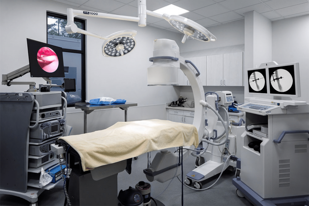
Endoscopy is an advanced, minimally invasive diagnostic procedure that uses a small, flexible camera to visualize the interior of your pet’s body in real time. This invaluable tool allows veterinarians to examine the gastrointestinal tract, respiratory system, and other internal areas without the need for traditional surgery. Endoscopy is commonly used to diagnose conditions such as chronic vomiting or diarrhea, gastrointestinal obstructions, foreign bodies, ulcers, inflammation, and airway abnormalities. It also enables tissue sampling and foreign body removal with reduced discomfort, shorter recovery times, and less stress for your pet, helping ensure accurate diagnosis and effective treatment.

Video Otoscopy

Video Otoscopy is an advanced diagnostic and treatment tool that uses a high-definition camera to provide a detailed, magnified view of your pet’s ear canal and eardrum. This invaluable technology allows veterinarians to accurately diagnose ear infections, inflammation, foreign bodies, tumors, and abnormalities deep within the ear that cannot be seen with standard examination tools. Video otoscopy also enables thorough ear cleaning, targeted treatment, and precise sampling, often under controlled conditions, resulting in improved outcomes, reduced discomfort, and more effective long-term management of ear disease.

A Better Life for Your Pet Start Here